Quantitative Assessment: Range of Motion
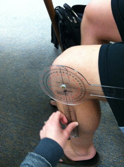
The most common and effective way to quantitatively test flexion and extension in the knee is to use a goniometer. A goniometer is an instrument used to measure the angles between two joints. The normative value for knee flexion is 130 degrees, while the normative value for knee extension is 0 degrees.
ACTIVE RANGE OF MOTION: The patient moves his/her body part of their own without help from the practitioner
PASSIVE RANGE OF MOTION: The practitioner moves the patient's body part.
RESISTIVE RANGE OF MOTION: The practitioner resists the patient's body part through full range of motion.
ACTIVE RANGE OF MOTION: The patient moves his/her body part of their own without help from the practitioner
PASSIVE RANGE OF MOTION: The practitioner moves the patient's body part.
RESISTIVE RANGE OF MOTION: The practitioner resists the patient's body part through full range of motion.
Qualitative assessment: Range of motion
Due to the fact that goniometric measurements are to be taken passively by the assessor, it is important to see how actively the patient can move the joint, in this case, the knee. These movements should be compared bilaterally, and changes in those comparisons should be noted.
Qualitative assessment: Manual muscle testing
Manual muscle testing is used to determine the strength of the muscles surrounding a joint. Due to the fact that the knee is only able to perform flexion and extension, those are the motions in which manual muscle testing will be used. Listed below is step-by-step instructions to perform manual muscle testing for the knee.
Manual Muscle Test: Hamstrings:
- Patient Position: Prone
- Starting Position: Knee Extended
- Stabilization: Femur
- Resistance: Distal Tibia
- Primary Mover(s): Biceps Femoris (Long Head and Short Head), Semimembranosus, Semitendinosus
- Secondary Movers: Gastrocnemius
- Substitution: Hip Flexion, Ankle Plantarflexion
- Comments: Internally Rotating the leg will emphasize contribution from the Popliteus, Semimembranosus, and Semitendinosus. Externally rotating the leg will emphasize contribution from the Biceps Femoris.
Manual Muscle Test: Quadriceps:
- Patient Position: Seated
- Starting Position: Knee flexed
- Stabilization: Distal Femur
- Palpation: Proximal to the Patella
- Resistance: Distal Tibia, proximal to the Ankle
- Primary Movers: Vastus Lateralis, Vastus Medialis, Vastus Intermedius, Rectus Femoris
- Substitution: Ankle Dorsiflexion, Hip Extension
Manual Muscle Test: Gluteus Maximus:
- Patient Position: Prone
- Starting Position: Knee is flexed to 90 degrees
- Stabilization: Posterior Pelvis
- Palpation: Posterior Thigh
- Resistance: Proximal to Popliteal Fossa
- Primary Movers: Gluteus Maximus
- Comments: Pain with the knee extended that decreases with the knee flexed implicates the Hamstrings. This can be confirmed by resisting knee flexion.
Manual Muscle Test: Isolating the Sartorius:
- Patient Position: Seated
- Starting Position: The heel of the leg being tested is positioned over the anterior Talocrural joint with the patient sitting over the edge of the table
- Stabilization: Distal Femur
- Palpation: Just inferior to the Anterior Superior Iliac Spine
- Resistance: Medial aspect of the distal Tibia and medial Ankle
- Primary Mover: Sartorius
- Secondary Movers: Hamstring group, Hip External Rotators, Gracilis, and Hip Flexors
- Substitution: Hip flexion without external rotation or abduction by the Rectus Femoris and/or Iliopsoas.
Manual Muscle Test: Tensor Fascia Latae:
- Patient Position: Side lying on unaffected side
- Starting position: Affected leg is extended and slightly abducted
- Stabilization: Iliac Crest
- Palpation: Lateral knee
- Primary Mover: Tensor Fascia Latae (Iliotibial Band)
Manual Muscle Test: Gracilis
- Patient Position: Side lying on affected side
- Starting Position: Affected leg is extended and adducted
- Stabilization: Iliac Crest of unaffected hip
- Palpation: Medial knee of the affected leg
-Primary Movers: Adductors including the Gracilis
Case scenario 1:
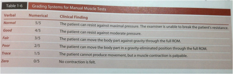
Functional assessment, bilaterally, indicates movements throughout AROM, PROM and RROM that are within normal limits. Manual muscle testing for the surrounding muscularture, listed above, produced a grade 5 throughout assessment.
case scenario 2:
Functional assessment in the form of active and passive range of motion was attempted for this athlete. The movements caused immense pain immediately following the hit, so movement was ceased. Girth measurements were taken for this athlete at the superior aspect of the patella, as well as 3' above, 6' above, 9' above and 12' above. These numbered were compared bilaterally in order to denote the amount of swelling present.
